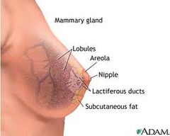
alveoli
smallest structures in a mammary gland
areola
darkened area surrounding nipple
colostrum
thin, yellow fluid, precursor of milk, secreted for a few days after birth
cooper ligaments
suspensory ligaments; fibrous bands extending from the inner breast surface to the chest wall muscles
fibroadenoma
benign breast mass
galactorrhea
persistent white discharge of milk between nursing sessions or after weaning
gynecomastia
excessive breast development in the male
intraductal papilloma
serosanguineous nipple discharge
inverted
nipples that are depressed or invaginated
lactiferous
conveying milk
mastitis
inflammation of the breast
Montgomery glands
sebaceous glands in the areola that secrete protective lipid during lactation; also called tubercles of Montgomery
Paget disease
intraductal carcinoma in the breast
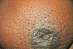
Peau d' orange
orange peel appearance of breast due to edema
retraction
dimple or pucker on the skin
striae
atrophic pink, purple, or white linear streaks on the breasts, associated with pregnancy, excessive weight gain, or rapid growth during adolescence
supernumerary nipple
minute extra nipple along the embryonic milk line
tail of spence
extension of breast tissue into the axilla
thelarche
beginning of prepubertal breast development
Appropriate history questions to ask regarding the breast examination.
-pain -lump -discharge -rash -swelling -trauma -history of breast disease - surgery or radiation -medications -patient-centered care -perform breast self-examination -last mammogram -tenderness, lump, or swelling -rash
Anatomy of the breast
Lactiferous duct; lactiferous sinus; lobule ; lobe; adipose tissue; cooper ligament; pectoralis major muscle
normal developmental stages of breast
1.preadolescent: there is only a small elevated nipple
2. breast bud stage: small mound of breast and nipple develops; the areola widens
3. the breast and areola enlarge; the nipple is flush with the breast surface
4. the areola and nipple form a secondary mound over the breast
5. mature breast: only the nipple protrudes; the areola is flush with the breast contour the areola may continue as a secondary mound in some normal woman
components of the breast examination
lie down. press the 3 middle fingers in a circular motion and use 3 levels of pressure. follow an up and down pattern.
sit up. examine underarm with arm slightly raised.
note surface changes with hands pushed on hips, shoulders hunched
points for self breast examination
-technique
-timing
-know what is normal for yourself
significance of a supernumerary nipple or breast
normal and common variation. an extra nipple along the embryonic "milk line" on the thorax or abdomen is a congenital finding. no associated glandular tissue. Tiny areola and nipple; distinguish from a mole.
difference between male and female examination procedures and findings
male exams are shorter; painless, firm retroareolar lump; less frequent signs of nipple discharge, ulceration, retraction, axially lymphadenopathy.
benign breast disease
-fibrocystic -multiple tender masses
-fibroadenoma- solitary nontender mass, solid, firm, rubbery
fibrocystic; multiple tender masses that occur with numerous symptoms and physical findings:
-swelling and tenderness -mastalgia -nodularity (lumpiness) - dominant lumps - nipple discharge -infections and inflammations
nodules are mobile, well demarcated, and feel rubbery like small water balloons
abscess
rare complication of generalized infection if untreated; a pocket of pus that feels hard, looks red, and is quite tender.
acute mastitis
uncommon; inflammatory mass before abscess formation. single quadrant. area is red, swollen, tender, very hot, and hard. may be from a plugged duct
breast cancer
solitary, unilateral, nontender mass. single focus in one area. solid, hard, dense, and fixed to underlying tissues or skin as cancer becomes invasive. borders are irregular, and poorly delineated. grows constantly. often painless
describe the characteristics to consider when a mass is noted in the breast
-location -size -shape - consistency -moveable - distinctness -nipple - note the skin over the lump -tenderness -lymphadenopathy
define gynecomastia
benign growth of this breast tissue; making it distinguishable from the other tissues in the chest wall. feels like a smooth, firm, movable disk. temporary. can also occur with use of anabolic steroids, some medications, cirrhosis, and other diseases.
describe screening mammography and clinical breast examination for the diagnosis of breast lesions
vertical strip palpitation - 3 different pressures
list the high risk and moderate risk factors that increase the usual risk for breast cancer
tumor suppressor genes termed BRCA 1 and BRCA2; inherit a mutation.
long term use of hormone replacement therapy increase risk
alcohol
breastfeeding is a risk reducer
What are the reservoirs for storing milk in the breast?
lactiferous sinuses
What is the most common site of breast tumors. what quadrant
upper outer quadrant
during a visit for a school physical, the 13 year old girl being examined questions the asymmetry of her breasts. what is the nurses best response?
one breast normally may grow faster than the other during development
when teaching the breast self-examination you would inform the woman that the best time to conduct breast self examination is:
Right after the menstrual period day 4 to 7 of the cycle; when the breasts are the smallest and least congested
you are providing health promotion teaching for a 40 year old woman. what is the current recommendation for women 40 years of age and older for breast cancer screening with mammography?
every year
you are going to inspect a female patients breasts for retraction .the best position for this part of the exam is?
sitting with hand pushing onto hips
A bimanual technique may be the preferred approach for a woman who is:
with pendulous breasts
During the examination of a 70 year old man you note gynecomastia you would:
review the medications for drugs that have gynecomastia as a side effect
during a breast exam you detect a mass. which of the following is most consistent with cancer rather than benign breast disease?
irregular, poorly defined, fixed
during the exam of the breasts of a pregnant woman, you would expect to find
blue vascular pattern over both breasts
which woman should not be referred to a physician for further evaluation?
a 25 year old with asymmetric breasts and inversion of nipples since adolescence
any lump found in the breast should be referred for further evaluation. a benign lesion will usually have 3 of the following characteristics. which one is characteristic of a malignant lesion?
irregular shape
gynecomastia is:
enlargement of the male breast
which is the first physical change associated with puberty in girls?
breast bud development
during the exam of a 30 year odl woman, she asks about the 2 large moles that are below her breast. after examining the area, how would you respond?
this is a normal finding of supernumerary nipples that are developed
colostrum
rich with antibodies that protect the newborn against infection. first few days after delivery
neonatal breast enlarge and visible from maternal estrogen crossing placenta and producing white discharge called :
Witch's milk
breast dimpling
shallow dimple also called skin tether shown as a skin retraction.
cancer causes firbrosis; which contracts the suspensory ligaments.
peau d' orange
edema of the breast; lymphatic obstruction
fixation
asymmetry, distortion, or decreased mobility with the elevated arm maneuver. cancer can fix the breast to the underlying pectoral muscles.
deviation in nipple pointing
underlying cancer causese fibrosis in the mammary ducts, which pulls the nipple angle toward it.
paget disease
intraductal carcinoma - early lesion has unilateral, clear, yellow discharge, and dry scaling crusts, spreasds outward to areola with erythematous halo on areola and crusted, eczematous, retracted nipple


