agnosia
loss of ability to recognize importance of sensory impressions
agraphia
loss of ability to express thoughts in writing
amensia
loss of memory
analogesia
loss of pain sensation
aphasia
loss of power of expression by speech, writing, or signs, or loss of comprehension of spoken or written language.
apraxia
loss of ability to perform purposeful movements in the absence of sensory or motor damage such as the inability to use objects correctly.
ataxia
inability to perform coordinated movements.
athetosis
bizarre, slow, twisting, writhing movement, resembling a snake or worm
chorea
sudden, rapid, jerky, purposeless movement involving limbs, trunk, or face.
clonus
rapidly alternating involuntary contraction and relaxation of a muscle in response to sudden stretch
coma
state of profound unconsciousness from which person cannot be aroused.
decerebrate rigidity
arms stiffly extended, adducted, internally rotated; legs stiffly extended, plantar-flexed.
decorticate rigidity
arms adducted and flexed, wrists and fingers flexed; legs extended, internally rotated, plantar-flexed.
dysarthria
imperfect articulation of speech due to problems of muscular control resulting from central or peripheral nervous system damage.
Dysphagia
impairment in speech consisting of lack of coordination and inability to arrange words in their proper order.
extinction
disappearance of conditioned response
fasciculation
rapid continuous twitching of resting muscle without movement of limbs
flaccidity
loss of muscle tone, limp
graphesthesia
ability to "read" a number by having it traced on the skin
hemiplegia
loss of motor power, paralysis, on one side of the body, usually caused by a stroke; paralysis occurs on side opposite the lesion.
lower motor neuron
motor neuron in the peripheral nervous system with its nerve fiber extending out to the muscle and only its cell body in the central nervous system
myoclonus
rapid sudden jerk of a muscle
nuchal rigidity
stiffness in cervical neck area
nystagmus
back and forth oscillation of the eyes
opisthotonos
prolonged arching of back, with head and heels bent backward, and meningeal irritation
paralysis
decreased or loss of motor function due to problem with motor nerve or muscle fibers
paraplegia
impairment or loss of motor and/or sensory function in the lower half of the body
paresthesia
abnormal sensation, burning, numbness, tingling, prickling, crawling, skin sensation
point localization
ability of the person to discriminate exactly where on the body the skin has been touched
proprioception
sensory information concerning body movements and position of the body in space
spasticity
continuous resistance to stretching by a muscle due to abnormally increased tension, with increased deep tendon reflexes
stereognosis
ability to recognize objects by feeling their form, size and weight while the eyes are closed
cerebral cortex
Is the center for humans highest functions governing thought, memory, reasoning, sensation and voluntary movement.
Frontal lobe
Area Concerned with personality, behavior, emotions, and intellectual function.
Parietal lobe
Postcentral gyrus: Primary center for sensation
Temporal lobe
Primary auditory receptor center with functions in hearing, taste, and smell
Wernicke's area
Associated with language comprehension. When damaged, receptive aphasia results meaning the person hears sound, but it has no meaning.
Ex. Like hearing a foreign language
-Broca's area
In frontal lobe, mediates motor speech. When injured, expressive aphasia results meaning the person cannot talk.
Ex. Can understand language and know what he wants to say but can't produce the language.
Basal ganglia
large bands of gray matter buried deep within the 2 cerebral hemispheres that form the subcortical associated motor system. They help to initiate and coordinate movement and control automatic associated movements of the body.
Ex. Arm swing alternating with legs while walking.
Thalamus
Main relay station where sensory pathways of the spinal cord, cerebellum, and brainstem form synapses on their way to the cerebral cortex.
synapses
sites of contact between two neurons
Hypothalamus
Major respiratory center with basic vital functions: temp, appetite, sex drive, heart rate, and blood pressure control, sleep center, anterior and posterior pituitary gland regulator, and coordinator of autonomic nervous system activity and stress response.
Cerebellum
Coiled structure under occipital lobe that is concerned with motor coordination of voluntary movements, equilibrium and muscle tone. Does NOT initiate movement but coordinates and smoothes.
Ex. the complex and quick coordination of many different muscles needed in playing piano, swimming, juggling
equilibrium
postural balance of body
Midbrain
most anterior part of brainstem that still has the basic tubular structure of the spinal cord; merges into the thalamus and hypothalamus; contains motor neurons and tracts.
Pons
Enlarged area of brainstem, containing ascending sensory and descending motor tracts. Includes the pneuotaxic and apneustic respiratory centers that coordinate with the main respiratory center in the medulla.
Medulla
Continuation of spinal cord in the brain that contains all ascending and descending fiber tracts; has vital autonomic centers of respiration, heart GI function, as well as nuclei for cranial nerves VIII - XII. Pyramidal decussation happens here: crossing of motor fibers.
Spinal cord
Long, cylindric structure of nervous tissue; occupies upper two-thirds of the vertebral canal from the medulla to lumbar vertebrae L1-L2. Its white matter is bundles of myelinated axons that form main highway for ascending and descending fiber tracts that connect brain to spinal nerves; it mediates reflexes of posture control, urination, pain response; its nerve cell bodies, or gray matter, are arranged in a butterfly shape with anterior and posterior "horns".
2. List the primary sensations mediated by the 2 major sensory pathways of the CNS.
The spinothalamic tract transmits sensations of pain, temperature, and crude or light touch.
The posterior (dorsal) column allows position (proprioception) without looking resulting in knowledge of where your body parts are in space and in relation to each other.
Vibration is the feeling of vibrating objects.
Stereognosis is finely localized touch where you can identify familiar objects by touch (travel to thalamus).
3. Describe 3 major motor pathways in the CNS, including the type of movements mediated by each.
Corticospinal or pyramidal tract: permits humans to have very skilled and purposeful movements.
Extrapyramidal tract: originates in the motor cortex, basal ganglia brainstem, and spinal cord; these subcortical motor fibers maintain muscle tone and control body movements, especially gross automatic movements such as walking.
Cerebellar system: coordinates movement, maintains equilibrium, and helps maintain posture; receives information about the position of muscles and joints, the body's equilibrium, and what kind of motor messages are being sent from the cortex to the muscles on feedback pathways.
4. Differentiate an upper motor neuron from a lower motor neuron.
Upper motor neuron:
located in the CNS, they are a complex of descending motor fibers that can influence or modify the lower motor neurons. They convey impulses from motor areas of the cerebral cortex to the lower motor neurons in the anterior horn.
Lower motor neuron:
located in the PNS, the cell body of the lower motor neuron is located in the anterior gray column of the spinal cord, with nerve fiber extending from to the muscle. Many neural signals spark here providing the final direct contact with muscles.
5. List the 5 components of a deep tendon reflex arc.
1. an intact sensory nerve (afferent)
2. a functional synapse in the cord
3. an intact motor nerve fiber (efferent)
4. neuromuscular junction
5. a competent muscle
6. List the major symptom areas to assess when collecting a health history for the neurologic system.
Assess: headache, head injury, dizziness/vertigo, seizures, tremors, weakness, incoordination, numbness or tingling, difficulty swallowing, difficulty swallowing, difficulty speaking, significant past history (stroke, spinal cord injury, meningitis), and environmental/occupational hazards
7. List and describe 3 tests of cerebellar function.
Observe as the patient walks 10 to 20 feet, turns, and returns to the starting point. Observe a heel-to-toe walk and a walk on tip-toes to assess balance. The normal gait should be smooth, rhythmic, and effortless with a coordinated opposing arm swing and smooth turns. An uncoordinated or unsteady gait are abnormal.
The Romberg Test has the patient stand up with feet together and arms at the sides. Once in a stable position, ask them to close the eyes and to hold the position.
Test the shallow knee bend or to hop in place, first on one leg, then the other followed by rapid alternating movements and pats on the knees with hands.
The finger-to-finger test asks the client to use their index finger to touch your finger then or his own nose as the position of your finger changes.
The finger-to nose test ask the patient to touch the tip of his nose with each index finger while alternating hands and increasing speed.
The heel-to-shin test places the heel on the opposite knee followed by running it down the shin from the knee to the ankle.
8. Describe the method of testing the sensory system for pain, temperature, touch, vibration, and position.
Pain: break a tongue blade for a "sharp" and "dull" side. Lightly apply the sharp point or the dull end to the person's body in a random, unpredictable order. Ask the person to say "sharp" or "dull" depending on the sensation felt.
--Abnormal findings:
---hypoalgesia- decreased pain sensation
---hyperalgesia-increased pain sensation
Temperature: place the flat side of the tuning fork on the skin; its metal always feels cool.
Light touch: wisp of cotton over the arms, forearms, hands, chest, thighs and legs. Ask the person to say "now" or "yes" when touch is felt.
---abnormal findings:
---hypoesthesia- decreased touch sensation
---anesthesia- absent touch sensation
---hyperesthesia- increased touch
9. Define the 4-point grading scale for deep tendon reflexes.
4+ very brisk, hyperactive with clonus, indicative of disease
3+ brisker than average, may indicate disease, probably normal
2+ average, normal
1+ diminished, low normal, or occurs only with reinforcement
0 no response
10. State the vertebral level whose intactness is accessed when eliciting each of these reflexes:
Biceps reflex: C5-C6 - blow to biceps tendon
triceps reflex: (C5-C6)--blow to biceps tendon
quadriceps reflex: (C2-C4)--(knee jerk) strike tendon just below patella
Achilles reflex: (C5-S2): ankle jerk
11. List the components of the neurologic recheck examination that are performed routinely on hospitalized patients monitored for neurologic deficit.
Level of consciousness: check orientation to the place and time; note the quality and the content of the verbal response: articulation, fluency, manner of thinking, and any deficit in language comprehension or production.
Motor function: giving the person specific commands: lift the eyebrows, frown, bare teeth; check upper arm strength by checking hand grasps; ask person to lift hand up; check lower by having them lift their leg.
Pupillary response: note the size, shape, and symmetry of both pupils: shine a light into each pupil and note the direct and consensual light reflex. Both pupils should constrict briskly.
Vital signs: blood pressure is notoriously an unreliable parameters of CNS deficit.
12. List the 3 areas of assessment on the Glasgow Coma Scale.
Best eye: opening response: spontaneously, to speech, to pain, no response
Best motor response: obeys, verbal command, localizes pain, flexion-withdrawal, flexion-abnormal, extension-abnormal, no response.
Best verbal response: conversation-confused, speech, sounds, no response
A fully normal person has a score of 15. 7 or less reflects a coma.

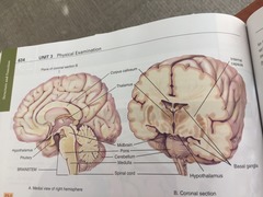
12a. Fill in the labels indicated in the following illustrations (the brain).
Plane of coronal section B
Hypothalamus
Pituitary
Brainstem
Spinal cord
Medulla
Cerebellum
pona
thalamus
corpus callosum
internal capsule
basil ganglia
hypothalamus

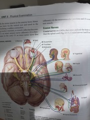
12b. Fill in the name of each cranial nerve, and then write S for sensory, M for motor, or MX for mixed.
1. Olfactory
2. Optic
3. Oculomotor
4. Troclear
5. Trigeminal
6. Abducens
7. facial
8. Acoustic
9. Glossopharyngeal
10. vagus
11. Accessory
12. Hypoglossal
1. The medical record indicates that a person has an injury to Broca's area. When meeting this person, you expect:
A. difficulty speaking
2. The control of body temperature is located in the:
D. hypothalamus.
3. To test for stereognosis, you would:
C. place a coin in the person's hand and ask them to identify it.
4. During the examination of an infant, use a cotton-tipped applicator to stimulate the anal sphincter. The absence of a response suggests a lesion of:
C. S2
5. During a neurologic examination, the tendon reflex fails to appear. Before striking the tendon again, you use the technique of:
B. reinforcement
6. Cerebellar function is assessed by which of the following:
C. coordination- hopping on one foot.
7. To elicit the Babinski reflex:
B. Stroke the lateral aspect of the sole of the foot from heel to across the ball.
8. A positive Babinski sign is:
A. Dorsiflexion of the big toe and fanning of all toes
9. The cremasteric response:
B. Is positive when the ipsilateral testicle elevates on stroking of the inner aspect of the thigh.
10. Senile tremors may resemble Parkinsonism, except that senile tremors do not include:
B. Rigidity and weakness of voluntary movement.
11. Patients who have Parkinson's disease usually have which of the following characteristic styles of speech?
C. Slow, monotonous.
12. The Glasgow Coma Scale, GCS, is divided into three areas; including:
B. Eye opening, motor response to stimuli, and verbal response.
13. The landau reflex in the infant is seen when:
D. The baby raises his head and arches the back, as in a swan dive.
14. A 65-year-old man has noticed a change in his personality and his ability to understand. He also cries and becomes angry very easily.
A. The cerebral lobe responsible for these behaviors is the frontal Lobe.
15. Olfactory:
F. Smell
16. Optic:
B. Vision
17. Oculomotor:
G. Extraocular movement, pupil constriction, down and inward movement of the eyes
18. Troclear:
K. Down and inward movement of the eye
19. Trigeminal:
H. Mastication and sensation of face, scalp, cornea
20. Abducens:
C. Lateral movement of the eye
21. Facial:
L. Tasting anterior 2/3s of tongue, closing eyes.
22. Acustic:
D. Hearing and equilibrium
23. Glossopharyngeal:
I. Phonation, swallowing, tasting posterior third of tongue.
24. Vagus:
E. Talking, swallowing, and sensory information from pharynx and carotid sinus.
25. Spinal:
J. Movement of trapezius and sternomastoid muscle
26. Hypoglossal:
A. movement of the tongue
