allen test
test that determines the patency of the radial and ulnar arteries by compressing one artery site and observing return of skin color as evidence of patency of the outer artery
aneurysm
defect of sac formed by dilation in artery wall due to atherosclerosis, trauma, or congenital defect
arrhythmia
variation from the hearts normal rhythm
arteriosclerosis
thickening and loss of elasticity of the arterial walls
atherosclerosis
plaques of fatty deposits formed in the inner layer, intima, of the arteries
bradycardia
slow heart rate, less than 50 beats per minute in the adult
bruit
blowing, swooshing sound heard through a stethoscope when an artery is partially occluded
cyanosis
dusky blue mottling of the skin and mucous membrane due to excessive amount of reduced hemoglobin in the blood
diastole
in hearts filling phase
ischemia
deficiency of arterial blood to a body part due to constriction of obstruction of a blood vessel
lymph nodes
small oval clumps of lymphatic tissue located at grouped intervals along lymphatic vessels
lymphedema
swelling of extremity due to obstructed lymph channel, nonpitting
pitting edema
indentation left after examiner depresses the skin over swollen edematous tissue
profile sign
viewing the finger from the side to detect early clubbing
pulse
pressure wave created by each heartbeat, palpable at body sites where the artery lies close to the skin and over a bone
pulsus bigeminus
irregular rhythm; every other beat is premature; premature beats have weakened amphlitude
pulsus paradoxus
beats have weaker amplitude with respiratory inspiration, stronger with expiration
systole
the hearts pumping phase
tachycardia
rapid heart rate, more than 95 beats per minute in the adult
thrombophlebitis
inflammation of the vein associated with thrombus formation
ulcer
open skin lesion extending into dermis, with sloughing of necrotic inflammatory tissue
varicose veins
dilated tortuous veins with incompetent valves
1. Describe the structure and function of arteries and veins.
The heart pumps freshly oxygenated blood through the arteries to all body tissues. Artery walls are strong, tough, and tense to withstand pressure demands; contain elastic fibers, which allow their walls to stretch with systole and recoil with diastole.
Veins are often closer to the skin and contain valves to help keep blood flowing toward the heart, while arteries carry blood away from the heart. Most veins carry deoxygenated blood from the tissues back to the heart; exceptions are the pulmonary and umbilical veins that carry oxygenated blood to the heart. Arteries are more muscular than veins.
2. List the pulse sites accessible to examination.
Temporal artery, carotid artery, brachial artery, femoral artery, popliteal artery, dorsalis pedis, posterior tibial.
3. Describe 3 mechanisms that help return venous blood to the heart.
1. The contracting skeletal muscles that milk the blood proximally, back toward the heart
2. The pressure gradient caused by breathing, in which inspiration makes the thoracic pressure decrease and the abdominal pressure increase
3. The intraluminal valves, which ensure unidirectional flow.
4. Define the term capacitance vessels, and explain its significance.
Veins have a larger diameter and are more distensible; they can expand and hold more blood when blood volume increases. This is a compensatory mechanism to reduce stress on the heart; this ability to stretch, veins are called capacitance vessels.
5. List the risk factors of venous stasis.
Elderly, diabetes, obesity, peripheral vascular disease, pregnancy, smoking, varicose veins, inactivity
6. Describe the function of the lymphatic system.
Retrieves excess fluid from the tissue spaces and returns it to the bloodstream. During circulation of the blood, somewhat more fluid leaves the capillaries than the veins can absorb. Without lymphatic drainage, fluid would build up in the interstitial spaces and produce edema.
7. Describe the function of the lymph nodes.
Nodes filter the fluid before it is returned to the bloodstream and filter out the micro-organisms that could be harmful to the body.
8. Name the related organs in the lymphatic system.
- Spleen;
(1) to destroy old red blood cells
(2) to produce antibodies
(3) to store red blood cells
(4) to filter micro-organisms from the blood
- Tonsils; respond to local inflammation
- Thymus; important in developing the T lymphocytes if the immune in children
9. List the symptom areas to address during history taking of the peripheral vascular system.
- Leg pain or cramps
- Skin changes on arms or legs
- Swelling in the arms or legs
- Lymph node enlargement
- Medication
10. Fill in the grading scale for assessing the force of an arterial pulse: 0 _____; 1+ _______; 2+_______; 3+_______.
Fill in the grading scale for assessing the force of an arterial pulse: • 0 = absent
• 1+ , weak
• 2+, normal
• 3+, increased,
• 4+ full, bounding
11. List the skin characteristics expected with arterial insufficiency to the lower legs.
Malnutrition, pallor and coolness; thin, shiny atrophic skin, thick-ridged nails, loss of hair, ulcers, gangrene
12. Compare the characteristics of leg ulcers associated with arterial insufficiency with ulcers with venous insufficiency.
Arterial deficit ulcers occur on tips of toes, metatarsal heads, and lateral malleoli.
Venus deficit is in the medial malleolus and lower leg; uneven edges superficial ulcer base, granulation tissue, beefy red to yellow, bleeding, may or may not be painful.
13. Fill in the description of the grading scale for pitting edema: 1+, 2+, 3+, 4+.
• 1+ Mild pitting, slight indentation, no perceptible swelling of the leg
• 2+ Moderate pitting, indentation, subsides rapidly
• 3+ Deep pitting, indentation remains for a short time, leg looks swollen
• 4+ Very deep pitting, indentation lasts a long time, leg is very swollen
14. Describe the technique for using the Doppler ultrasonic probe to detect peripheral pulses.
-Position person supine, with legs externally rotated so you can reach the medial ankles easily
-Place a drop of coupling gel on the end of the handheld transducer
-Place the transducer over a pulse site, swivelled at a 45o angle.
-Apply very light pressure, locate the pulse site by the swishing, whooshing sound
15. Raynaud phenomenon has associated progressive tricolor changes of the skin from blue to white and then to
red. State the mechanism for each of these color changes.
(1) white (pallor) in top figure from arteriospasm and resulting deficit in supply;
(2) blue (cyanosis) in lower figure from slight relaxation of the spasm that allows a slow trickle of blood through the capillaries and increased oxygen extraction of hemoglobin;
(3) finally, red (rubor) in heel of hand due to return of blood into the dilated capillary bed or reactive hyperemia.
May have cold, numbness, or pain along with pallor or cyanosis stage; then burning, throbbing pain, swelling along with rubor. Lasts minutes to hours; occurs bilaterally. Several drugs predispose to the episodes, and smoking can increase the symptoms.
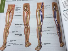
15a. Fill in the labels indicated in the following arteries and name the pulse sites.
abdominal aorta
common iliac artery
external iliac artery
femoral pulse site
popliteal artery
popliteal pulse site
anterior tibial artery
posterior tibial artery
dorsalis pedis artery
dorsal arch
dorsal pedis pulse site
posterior tibial pulse site
1. A function of the venous system includes:
A. Holding more blood when blood volume increases
Spleen, tonsils, and thymus
organs that aid the lymphatic system
During pregnancy: dependent edema, varicosities in the legs, and hemorrhoids.
"The symptoms are caused by the pressure of the growing uterus on the veins. They are usual conditions of pregnancy".
pulse amplitude of 3+
Is increased and full
Epitrochlear node
5. Inspection of a person's right hand reveals a red swollen area. To further assess for infection, you would palpate the:
screening for deep vein thrombosis
Measure the wildest point with a tape measure
If you are unable to palpate the popliteal pulse.
Proceed with the examination. It is often impossible to palpate this pulse.
document 4+ edema of the right.
D. Very deep pitting, indentation lasts a long time.
arterial deficit in the lower extremities. After raising the legs 12 inches off the table and then having the person sit up and dangle the leg, the color should return in.
After raising the legs 12 inches off the table and then having the person sit up and dangle the leg, the color should return in 10 seconds or less.
characteristic sign of varicose veins
in lower extremities; dilated, tortuous superficial bluish vessels.
peripheral arterial insufficiency include:
Atrophic skin changes; thin, shiny skin with loss of hair
Intermittent claudication includes
Muscular pain brought on by exercise
venous ulcer risk factor
obesity
Arterioscerosis cause
Loss of elasticity of the walls of blood vessels
Raynaud phenomenon occurs:
In hands and feet as a result of exposure to cold, vibration, and stress


